Home
/ Pelvic Anatomy Xray - 1 - Systematically examine all bony structures of the pelvis and femurs for symmetry, cortical breaks and joint spaces (sacroiliac, hip and.
Pelvic Anatomy Xray - 1 - Systematically examine all bony structures of the pelvis and femurs for symmetry, cortical breaks and joint spaces (sacroiliac, hip and.
Pelvic Anatomy Xray - 1 - Systematically examine all bony structures of the pelvis and femurs for symmetry, cortical breaks and joint spaces (sacroiliac, hip and.. Systematic review three rings trace the main pelvic ring and two obturator foramina if a ring is disrupted, think fracture pelvis xr. If line disruption, think fractured proximal femur. Anatomy pelvis at university of kansas medical center the sacroiliac joints should be symmetrical joint space range 2 4 mm. Pelvic floor anatomy is complex and is being unraveled by means of magnetic resonance mr imaging. Each innominate bone is composed of three parts, which fuse at the acetabulum.
Learn vocabulary, terms and more with flashcards only rub 220.84/month. If either joint space is widened think main pelvic ring fracture. The main purposes of the pelvic girdle are to support and protect the abdominal and pelvic organs, and to connect the trunk and lower limbs. For more anatomy content please follow us and visit our website: Anatomy of the pelvis the pelvis is a ring of bones situated between the spine and the legs.
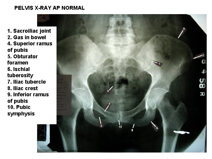
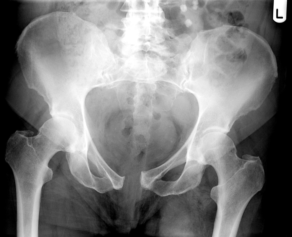
Abdomen And Pelvis Radiographs And Images Xrays And from slidetodoc.com An x ray of the pelvis focuses specifically on the area between your hips that holds many of your reproductive. The bony pelvis, the pelvic cavity, the pelvic floor, and the perineum. Pelvis x ray anatomy in this image you will find the sacroiliac joint acetabular obturator foramina greater trochanter pubic symphysis femoral heads lesser trochanters in it. Human anatomy for muscle, reproductive, and skeleton. Pelvic ring formed from 2 innominate. Siu/icud consultation on urethral strictures: A pelvis x ray also known as a pelvis series or pelvis radiograph is a single x ray of the pelvis to include the iliac crests and pubic symphysis. Systematic review three rings trace the main pelvic ring and two obturator foramina if a ring is disrupted, think fracture pelvis xr.
If either joint space is widened think main pelvic ring fracture.
●to review pelvic sidewall anatomy including retroperitoneal spaces. Human anatomy for muscle, reproductive, and skeleton. The space or compartment surrounded by the pelvic girdle (bony pelvis). This is an online quiz called elbow xray anatomy. However, infectious, inflammatory, neoplastic, and traumatic processes frequently. The cortex of femoral head, neck, greater, and lesser trochanter should be smooth with normal trabecular pattern on ap and lateral. The bony pelvis, the pelvic cavity, the pelvic floor, and the perineum. The pelvic spine consists of. Our latest youtube film is ready to run. The sagittal (longitudinal) image of the female pelvis shows anatomical structures. The judet view is comprised of two projections first the iliac oblique for assessment of the posterior column and anterior wall of the acetabulum. Surgical pelvic anatomy in gynecologic oncology. If either joint space is widened think main pelvic ring fracture.
In an adult, the innominate bones consist of the fused ilium, ischium, and pubis (figure 1). Pelvic ring formed from 2 innominate. Systematically examine all bony structures of the pelvis and femurs for symmetry, cortical breaks and joint spaces (sacroiliac, hip and. The bony pelvis, the pelvic cavity, the pelvic floor, and the perineum. Use the mouse scroll wheel to move the images up and down alternatively use the tiny arrows (>>) on both side of the image to move the images.>>) on both side of the image to move the images.
The Pelvis And Hip from www.imageinterpretation.co.uk To review pelvic sidewall anatomy including retroperitoneal spaces. The anorectal hiatus is the only opening in the pelvic diaphragm. The main purposes of the pelvic girdle are to support and protect the abdominal and pelvic organs, and to connect the trunk and lower limbs. In an adult, the innominate bones consist of the fused ilium, ischium, and pubis (figure 1). Pelvis x ray anatomy in this image you will find the sacroiliac joint acetabular obturator foramina greater trochanter pubic symphysis femoral heads lesser trochanters in it. Anatomy pelvis at university of kansas medical center the sacroiliac joints should be symmetrical joint space range 2 4 mm. Pelvic floor anatomy is complex and is being unraveled by means of magnetic resonance mr imaging. Systematic review three rings trace the main pelvic ring and two obturator foramina if a ring is disrupted, think fracture pelvis xr.
An x ray of the pelvis focuses specifically on the area between your hips that holds many of your reproductive.
An x ray of the pelvis focuses specifically on the area between your hips that holds many of your reproductive. Hover on/off image to show/hide findings. Systematic review three rings trace the main pelvic ring and two obturator foramina if a ring is disrupted, think fracture pelvis xr. This is an online quiz called elbow xray anatomy. The cortex of femoral head, neck, greater, and lesser trochanter should be smooth with normal trabecular pattern on ap and lateral. It is difficult to identify normal peritoneal folds and ligaments at imaging. Annotated case courtesy of dr matthew lukies, radiopaedia.org, 51247. Although ultrasound images are another modality typically used for imaging the pelvic region, excluding ultrasound from the tutorial was a pedagogical decision, as the tutorial targets novice learners. Hemi pelvis anatomy normal ap. It includes several structures : Click here to load quiz. The coccyx, commonly referred to as the tailbone, is the smallest of the pelvic bones, and sits inferiorly to the. Your pelvis is made up of three bones, the ilium, ischium, and.
The muscle originates from the body of the pubis and attaches to the pectineal line and proximal part of the linea aspera of femur. Hemi pelvis anatomy normal ap. The judet view is comprised of two projections first the iliac oblique for assessment of the posterior column and anterior wall of the acetabulum. Anatomy pelvis at university of kansas medical center the sacroiliac joints should be symmetrical joint space range 2 4 mm. If either joint space is widened think main pelvic ring fracture.
Plain Radiographic Evaluation Of The Hip Musculoskeletal Key from musculoskeletalkey.com Tap on/off image to show/hide findings. Angiography invasive angiography is the gold standard modality for assessing pelvic vasculature 3. The cortex of femoral head, neck, greater, and lesser trochanter should be smooth with normal trabecular pattern on ap and lateral. Surgical pelvic anatomy in gynecologic oncology. If either joint space is widened think main pelvic ring fracture. The coccyx, commonly referred to as the tailbone, is the smallest of the pelvic bones, and sits inferiorly to the. A pelvis x ray also known as a pelvis series or pelvis radiograph is a single x ray of the pelvis to include the iliac crests and pubic symphysis. Click image to align with top of page.
Pelvic ring formed from 2 innominate.
Hemi pelvis anatomy normal ap. If line disruption, think fractured proximal femur. The pelvic spine consists of. Branches of the internal iliac artery. The bony pelvis, the pelvic cavity, the pelvic floor, and the perineum. Hemi pelvis anatomy normal ap. Pelvic anatomy knowledge, and on participant confidence with imaging in clinical situations. The cortex of femoral head, neck, greater, and lesser trochanter should be smooth with normal trabecular pattern on ap and lateral. An x ray of the pelvis focuses specifically on the area between your hips that holds many of your reproductive. Annotated case courtesy of dr phillip marsh, radiopaedia.org, rid: However, infectious, inflammatory, neoplastic, and traumatic processes frequently. It is subdivided into the greater pelvis and lesser pelvis. Über 7 millionen englischsprachige bücher.
This is an online quiz called elbow xray anatomy pelvic anatomy. Pelvic anatomy knowledge, and on participant confidence with imaging in clinical situations.